ملف:Schematic representation of the hypothalamic nuclei.png

حجم هذه المعاينة: 800 × 342 بكسل. الأبعاد الأخرى: 320 × 137 بكسل | 640 × 274 بكسل | 1٬276 × 546 بكسل.
الملف الأصلي (1٬276 × 546 بكسل حجم الملف: 539 كيلوبايت، نوع MIME: image/png)
| هذا ملف من ويكيميديا كومنز. معلومات من صفحة وصفه مبينة في الأسفل. كومنز مستودع ملفات ميديا ذو رخصة حرة. |
ملخص
| الوصف |
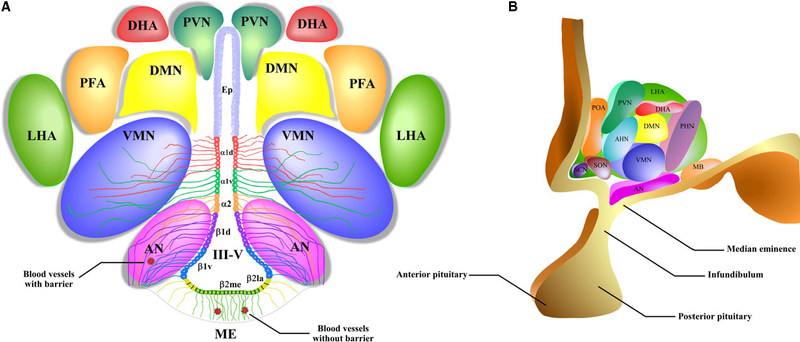
English: A schematic representation of the hypothalamic nuclei and the distribution of tanycytes over the wall of the third ventricle (III-V). (A) Coronal view of the approximate location of the hypothalamic nuclei and tanycytes. Ciliated ependymocytes (ep) line the dorsal wall of the III-V. The α1d-tanycytes (α1d) and α1v-tanycytes (α1v) have long projections that make contact with the neurons of the VMN. α2-tancycytes (α2) have projections to the AN and blood vessels. In a more ventral section of the III-V, the β1d-tanycytes (β1d) and β1v-tanycytes (β1v) make projections to the AN, making contact with orexigenic and anorexigenic neurons and blood vessels. In the floor of the III-V, the β2la-tanycytes (β2la) and β2me-tanycytes (β2me) are joined by tight junctions forming part of the median eminence (ME)-cerebrospinal fluid (CSF) barrier, and their projections make contact with the fenestrated blood vessels of the ME. (B) Sagittal view of the distribution of the hypothalamic nuclei. Ep: ependymocytes; AN: arcuate nucleus; VMN: ventromedial nucleus; DMN: dorsomedial nucleus; PVN: periventricular nucleus; DHA: dorsal hypothalamic area; PFA: perifornical area; LHA: lateral hypothalamic area; SCN: suprachiasmatic nucleus; SON: supraoptic nucleus; POA: preoptic area; MB: mammillary bodies; ME: median eminence; III-V: third ventricle. |
| التاريخ | |
| المصدر | Elizondo-Vega R, Cortes-Campos C, Barahona MJ, Oyarce KA, Carril CA, García-Robles MA. The role of tanycytes in hypothalamic glucosensing. Journal of Cellular and Molecular Medicine. 2015;19(7):1471-1482. doi:10.1111/jcmm.12590. https://www.ncbi.nlm.nih.gov/pmc/articles/PMC4511346/ |
| المؤلف | Roberto Elizondo-Vega, Christian Cortes-Campos, Maria J Barahona, Karina A Oyarce, Claudio A Carril, and Maria A García-Robles |

ترخيص
هذا الملف مرخص تحت ترخيص المشاع الإبداعي الدولية المُلزِمة بنسب العمل إلى مُؤلِّفه 4.0.
- يحقُّ لك:
- مشاركة العمل – نسخ العمل وتوزيعه وبثُّه
- إعادة إنتاج العمل – تعديل العمل
- حسب الشروط التالية:
- نسب العمل إلى مُؤَلِّفه – يلزم نسب العمل إلى مُؤَلِّفه بشكل مناسب وتوفير رابط للرخصة وتحديد ما إذا أجريت تغييرات. بالإمكان القيام بذلك بأية طريقة معقولة، ولكن ليس بأية طريقة تشير إلى أن المرخِّص يوافقك على الاستعمال.
تاريخ الملف
اضغط على زمن/تاريخ لرؤية الملف كما بدا في هذا الزمن.
| زمن/تاريخ | صورة مصغرة | الأبعاد | مستخدم | تعليق | |
|---|---|---|---|---|---|
| حالي | 19:41، 15 سبتمبر 2018 | | 1٬276 × 546 (539 كيلوبايت) | commonswiki>Was a bee | {{Information |Description={{en|1=A schematic representation of the hypothalamic nuclei and the distribution of tanycytes over the wall of the third ventricle (III-V). (A) Coronal view of the approximate location of the hypothalamic nuclei and tanycytes. Ciliated ependymocytes (ep) line the dorsal wall of the III-V. The α1d-tanycytes (α1d) and α1v-tanycytes (α1v) have long projections that make contact with the neurons of the VMN. α2-tancycytes (α2) have projections to the AN and blood vessel... |
استخدام الملف
ال1 ملف التالي مكررات لهذا الملف (المزيد من التفاصيل):
- ملف:Schematic representation of the hypothalamic nuclei.png من ويكيميديا كومنز
الصفحة التالية تستخدم هذا الملف: