|
تضامنًا مع حق الشعب الفلسطيني |
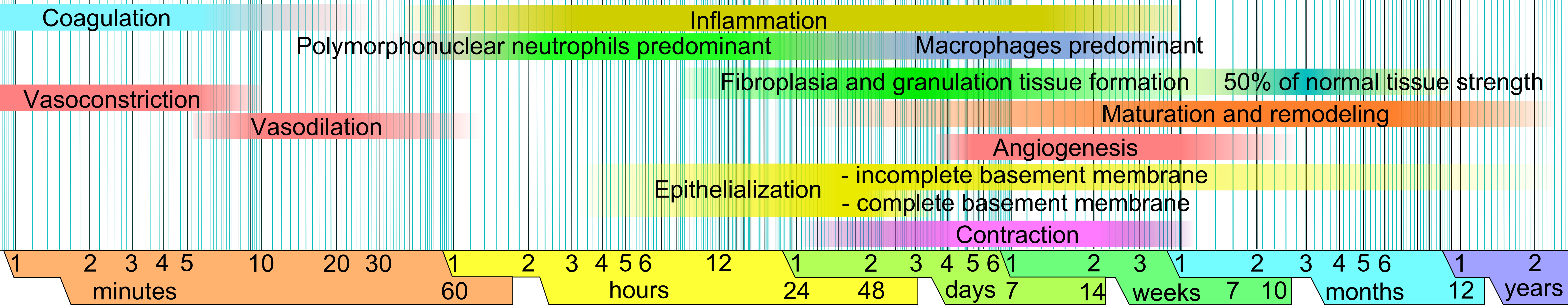
ملف:Wound healing phases.png
الملف الأصلي (6٬338 × 1٬236 بكسل حجم الملف: 859 كيلوبايت، نوع MIME: image/png)
| هذا ملف من ويكيميديا كومنز. معلومات من صفحة وصفه مبينة في الأسفل. كومنز مستودع ملفات ميديا ذو رخصة حرة. |
ملخص
| الوصف |
English: Phases of wound healing. Limits vary within faded intervals, mainly by wound size and healing conditions, but image does not include major impairments that cause chronic wounds. |
| التاريخ | |
| المصدر | عمل شخصي (from the template Logarithmic time scale - milliseconds to years.svg) |
| المؤلف |
When using this image in external works, it may be cited as:
or
|
| إصدارات أخرى | العربيَّة |
References
The direct URL link to this reference list is: http://commons.wikimedia.org/wiki/File:Wound_healing_phases.png#References
Inflammation and upper limit of beginning of maturation and remodeling, as well as its ending:
- worldwidewounds.com > Figure 3 - The time relationship between the different processes of wound healing. by Gregory S Schultz, Glenn Ladwig and Annette Wysocki - in turn adapted from Asmussen PD, Sollner B. Mechanism of wound healing. In: Wound Care. Tutorial Medical Series. Stuttgart: Hippokrates Verlag, 1993.
Lower limit of beginning of maturation and remodeling, and equivalent limit for fibroplasia and granulation tissue formation:
- Fig. 9-1. The cellular, biochemical, and mechanical phases of wound healing. Pollock, Raphael E.; F. Charles Brunicardi; Dana Lynne Andersen; Billiar, Timothy R.; Dunn, David; Hunter, John G.; Matthews, Jeffrey J. (٢٠٠٩) Schwartz's Principles of Surgery, Ninth Edition، McGraw-Hill Professional ISBN: 0-07-154769-X.
Vasoconstriction and vasodilation:
- Stadelmann W.K., Digenis A.G. and Tobin G.R. (1998). Physiology and healing dynamics of chronic cutaneous wounds. The American Journal of Surgery 176 (2): 26S-38S. PMID 9777970 Hamilton, Ont. B.C. Decker, Inc. Electronic book
Angiogenesis:
- Nguyen, D.T., Orgill D.P., Murphy G.F. (2009). Chapter 4: The Pathophysiologic Basis for Wound Healing and Cutaneous Regeneration. Biomaterials For Treating Skin Loss. CRC Press (US) & Woodhead Publishing (UK/Europe), Boca Raton/Cambridge, p. 25-57. (ISBN 978-1-4200-9989-9 Invalid ISBN, ISBN 978-1-84569-363-3)
Polymorphonuclear neutrophils and ending of fibroplasia and granulation tissue formation:
- de la Torre J., Sholar A. (2006). Wound healing: Chronic wounds. Emedicine.com. Accessed January 20, 2008. http://www.emedicine.com/plastic/topic477.htm
Macrophages:
- Expert Reviews in Molecular Medicine. (2003). The phases of cutaneous wound healing. 5: 1. Cambridge University Press. Accessed January 20, 2008. http://www-ermm.cbcu.cam.ac.uk/03005829a.pdf
Upper limit of beginning of fibroplasia and granulation tissue formation (collagen deposition), epithelialization and contraction:
- Romo T. and Pearson J.M. 2005. Wound Healing, Skin. Emedicine.com. Accessed December 27, 2006.
Additional note on contraction:
- Mulvaney M. and Harrington A. 1994. Chapter 7: Cutaneous trauma and its treatment. In, Textbook of Military Medicine: Military Dermatology. Office of the Surgeon General, Department of the Army. Virtual Naval Hospital Project. Accessed through web archive on September 15, 2007. https://web.archive.org/web/20031218072356/http://www.vnh.org/MilitaryDerm/Ch7.pdf
Percentage of normal tissue strength:
- Mercandetti M., Cohen A.J. (2005). Wound Healing: Healing and Repair. Emedicine.com. Accessed January 20, 2008. http://www.emedicine.com/plastic/topic411.htm

|
يُمثِّل: File:Wound healing phases.svg نسخةً شعاعيَّةً لهذه الصورة. ينبغي أن تستخدم الصورة الشعاعيَّة عندما تكون أعلى جودةً مِن الصورة النقطيَّة.
File:Wound healing phases.png → File:Wound healing phases.svg
للمزيد من المعلومات، راجع صفحة Help:SVG. |
ترخيص
| أنا، مالِك حقوق تأليف ونشر هذا العمل، أجعله في النِّطاق العامِّ، يسري هذا في أرجاء العالم كلِّه. في بعض البلدان، قد يكون هذا التَّرخيص غيرَ مُمكنٍ قانونيَّاً، في هذه الحالة: أمنح الجميع حق استخدام هذا العمل لأي غرض دون أي شرط ما لم يفرض القانون شروطًا إضافية. |
تاريخ الملف
اضغط على زمن/تاريخ لرؤية الملف كما بدا في هذا الزمن.
| زمن/تاريخ | صورة مصغرة | الأبعاد | مستخدم | تعليق | |
|---|---|---|---|---|---|
| حالي | 09:24، 18 يناير 2011 | 6٬338 × 1٬236 (859 كيلوبايت) | commonswiki>Mikael Häggström | Moved info in infobox to image page instead. |
استخدام الملف
ال1 ملف التالي مكررات لهذا الملف (المزيد من التفاصيل):
- ملف:Wound healing phases.png من ويكيميديا كومنز
الصفحة التالية تستخدم هذا الملف: