ملف:Functional magnetic resonance imaging.jpg

من أرابيكا، الموسوعة الحرة
اذهب إلى التنقل اذهب إلى البحث

Functional_magnetic_resonance_imaging.jpg(250 × 208 بكسل حجم الملف: 11 كيلوبايت، نوع MIME: image/jpeg)

ملخص

الوصف
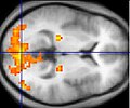
English: Sample fMRI data
This example of fMRI data shows regions of activation including primary visual cortex (V1, BA17), extrastriate visual cortex and lateral geniculate body in a comparison between a task involving a complex moving visual stimulus and rest condition (viewing a black screen). The activations (yellow-red) are shown (as is typical) against a background based on the average structural images from the subjects in the experiment.
التاريخ
المصدر
  • نُقِلت من en.wikipedia إلى كُومُنز .
  • عمل شخصي
المؤلف Washington irving في ويكيبيديا الإنجليزية

ترخيص

وُضِعت هذه الصُّورة في النِّطاق العامّ مِن قبل مُؤَلِّفها، Washington irving في مشروع Wikimedia Commons. ويسري هذا في جميع أَنحاء العالم.

إِذا لم يكن النِّطاق العام مُمكِناً مِن النَّاحية القانونيَّة:

يَمنح المُؤَلِّف Washington irving أَيَّ شخصٍ الحقَّ في استخدام هذا العمل لأَي سببٍ كان'، دون أَي شروطٍ من قبله، ويسري هذا ما لم يكن هُناك أَحكامٌ إِضافيَّة ينصُّ عليها القانون.

سجلُّ الرَّفع الأصيل

صفحة الوصف الأصلية كانت هنا، تشير جميع أسماء المستخدمين التالية إلى en.wikipedia.


Public-domain: copyright disclaimed Washington irving 07:49, 8 Mar 2004 (UTC)

الشروحات

أضف شرحاً من سطر واحد لما يُمثِّله هذا الملف

العناصر المصورة في هذا الملف

يُصوِّر

٨ مارس 2004

تاريخ الملف

اضغط على زمن/تاريخ لرؤية الملف كما بدا في هذا الزمن.

زمن/تاريخصورة مصغرةالأبعادمستخدمتعليق
حالي04:52، 9 ديسمبر 2004تصغير للنسخة بتاريخ 04:52، 9 ديسمبر 2004250 × 208 (11 كيلوبايت)commonswiki>SuperborsukSample fMRI data This example of fMRI data shows regions of activation including primary visual cortex (V1, BA17), extrastriate visual cortex and lateral geniculate body in a comparison between a task involving a complex moving visual stimulus and re

ال1 ملف التالي مكررات لهذا الملف (المزيد من التفاصيل):